Hello again! In previous posts I talked about how we were using a co-culture system to grow our leukemic cells. Although this is a great way to maintain cells, it can complicate some analyses where we cannot distinguish mouse (stroma) from human (AML) cells. We wanted to determine H3K27me3 levels (the mark that EZH2 is responsible for) in the AML patient cells we are working with. Usually this can be performed using the western blots. However, the antibody for the mark recognizes both human and mouse H3K27me3. To overcome this, we wanted to see if we could determine H3K27me3 levels using flow cytometry where we can distinguish the human cells from the mouse cells using the human CD45 marker. Experimental details can be found at Zenodo.
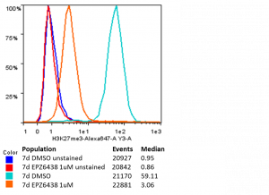
Figure 1: Histogram of H3K27me3 levels in CD45 positive OCI-AML-20 with (EPZ6438; red and orange) and without (DMSO; blue and light blue) 7 days of EZH2 inhibitor treatment. Number of events (cells) is displayed, as well as median fluorescent intensity (MFI) for cell populations. Unstained show almost no fluorescence. DMSO treated cells show a substantial fluorescence signal that is greatly reduced in the presence of EZH2 inhibitor.
This assay looks so clean… Congrats Genna!