Hello again,
Three weeks have now passed since my first blog, and the response has been overwhelming, who knew this kid with a love of dinosaurs could become an internet blogger. Your encouragement to make my t-Rex has motivated me to do just that, so let’s see how it all goes. Also fun fact, the biggest and oldest T-Rex was found in Canada, so looks like I am in right place for dinosaurs.
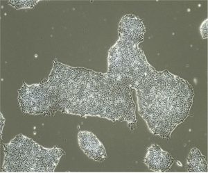
But, in the meantime, I do have a day, night and weekend job running a lab and academic drug discovery group, and so its time to discuss science. More specifically, what cells we work with in our work. Our bread and butter are the iPSC, short for induced pluripotent stem cells. Made from the blood of patients and healthy volunteers, when you look at them, they look like regular old cells, tightly packed into colonies, as they love being close to their fellow cells, problems arise when they want to move away and get some distance from their neighbors. Below are some images of what they look like, courtesy of Carol Chen and Narges Abdian, who help manage and oversee our catalog of stem cell lines in my group, along with Zhipeng You. As you can see, the cells are tightly packed, with each cell having the potential to grow into any cell type you want, all it needs is the right ingredients to direct it into the cell type of your choosing.
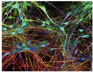
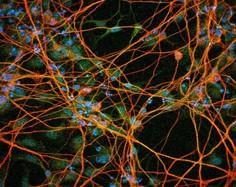
And what might these cells be, well we are interested in diseases of the brain, and in particular the cells lost during the progression of these degenerative disorders. This means our focus is on getting these stem cells to form neurons, of which everyone of us has over 100 billion. We can make many different types and over the coming months, I will be putting online our different SOPs from our group outlining how we make many of these different neurons. One of the main interests for our group is Parkinson’s disease, and the cells lost in this disease are dopaminergic neurons, making it our mission to understand why these neurons are being lost in the disease. Other cell types we make are motor neurons, sensory neurons and cortical neurons, all different types of neurons that are involved in different diseases, playing different roles. For those who wonder what they look like, here are some images below, staining for proteins that are specific for these neurons (Dopaminergic-Top; Cortical- Bottom). Images are courtesy of Carol Chen and Cecilia DeRocha.
In addition to neurons, we also make the support cells for these neurons, and one of these cells is astrocytes. This cell can be the good guy-bad guy and has been demonstrated to support neuronal survival, and on the flip side, to promote the loss of neurons. Courtesy of Vincent Soubannier, here are some images of what these cells look like in the dish.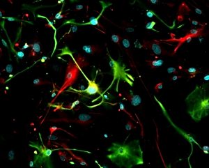
Now, I know, you are saying Tom, these neurons and astrocytes are beautiful, but they are flat, having been grown in 2D dishes. This isn’t like a brain; the brain is three dimensional and complex. And you know what, you would be right. Growing cells in 2D is artificial, but it lets us grow and image neurons and astrocytes for doing research on human cells that otherwise wouldn’t be accessible. But it wasn’t until the pioneering work by Dr Madeline Lancaster and others that the idea for making complex brain structures emerged. From pioneering work in her lab, she showed that neuronal organoids or “minibrains” could be formed in a dish. More complex then anything grown in 2D, these minibrains could be expanded, grown for several months, if not years, and show many characteristics of different regions within the human brain. Over 18 months ago, we turned our attention to minibrains and now after all this time, we can make three different types in our group: midbrain, cerebral and forebrain neuronal organoids or “minibrains”. You can learn more about this work through the enclosed link, with Dr Nguyen-Vi Mohamed showing how these are made in the lab.
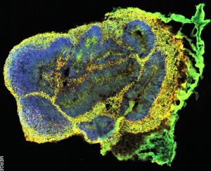
These can grow up to 4mm in size depending on the minibrain grown, and are comprised of neurons, astrocytes and many other cell types. They can be maintained for months, and our oldest minibrains were over 14 months old. Just as a preview of what they look like, below is a section through one of these minibrains at 30 days, courtesy of Dr Nguyen-Vi Mohamed.
With incubators packed with these little guys, we now have over 10,000 growing. These are probably the coolest things I have ever seen and am amazed at the things that can be done with them. Its such a new model, we are barely scratching the surface at what can be done, but I am looking forward to trying new things and seeing what others do. To end, I wanted to bring peoples attention to some remarkable work from from the Lancaster group published this week (I am like a fanboy of her work). They could connect a minibrain and muscle, with the minibrain taking control over the muscle and causing it to contract. This is just the tip of the iceberg and now I can’t wait to see what other structures we can connect to a minibrain. It won’t be long before we have little mini bodies on a dish, with minibrains connected to miniature versions of all the major organs in the body. But for now, we wait and see how the field progresses. For those wondering how we make minibrains, our methods paper is coming out in next week or so, and once its out, I will use this blog to cover the main points on how we make these organoid structures amongst other hot topics in the stem cell field. Next blog, I will be discuss how to thaw out a vial of iPSCs for all those new users wondering what I should do when I get a tube of these cells. Well don’t panic, I am here to help you. Feel free to connect with me on LinkedIn, if I don’t know you please at least introduce yourself and who you are. I also now on the twitter (@thomas.durcan), so you can follow me there too, I am a relative novice at this whole social media malarkey, so we shall see how that goes.