In a previous post, I determined a crystallization condition that produced nice 3D crystals of USP5 Zf-UBD that diffracted well. Rachel Harding, a postdoc at the SGC collected data images for these crystals and solved the structure using computational techniques. You can see the solved crystal structure and a more detailed analysis of the structure solution at Zenodo. Analysis of this structure reveals that the carboxyl group of glutathione (Figure 1), an additive used in the crystallization buffer, occupies the same position as the C-terminal ubiquitin COOH in the zinc finger binding pocket (Figure 2). Unfortunately, this means that the crystallization condition previously determined is not ideal for screening compounds as the binding pocket is not amenable to soaking with compounds since it is blocked by glutathione.
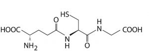
Figure 1. Glutathione structure
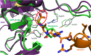
Figure 2. Superimposed structure of USP5 Zf-UBD (PDB code: 2G45 [purple]) and our new structure [green] showing that the carboxylate of glutathione [green] mimics the C-terminal extremity of ubiquitin [orange]
I’ll have to explore alternative crystallization conditions so the binding pocket is accessible for soaking with compounds!
One Reply to “Structural Analysis of USP5 Zf-UBD”