NSD3(PWWP1) and histone H3 interaction cellular assay

Background NSD3 is a member of the NSD (nuclear receptor SET domain- containing) family of H3K36 methyltransferases. It has two isoforms: long -1437 amino acid and short – 645 amino acids, which are derived from alternative splicing of exon 10.  In contrast to long isoform, the short isoform lacks the methyltransferase domain and it contains Read More …

Analysis of NSD3 Isoform Expression from TCGA-LUSC Data

There are two major NSD3 isoforms expressed, long (aa 1-1437) and short (aa 1-645, differing in sequence from 620-645). The short isoform lacks a methyltransferase domain and, perhaps surprisingly, is the form attributed to NSD3’s role in acute myeloid leukemia (AML) [1]. I’ve previously used the TCGA-LUSC data-set [2] to test for differential expression based on the Read More …

Purification of Recombinant Cas9

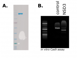
These days CRISPR gene editing needs little introduction. From humble origins as a bacterial defence system evolved to thwart viral invaders, CRISPR-based technologies have recently made headlines for their promise to revolutionize medicine. While there is a lot of excitement surrounding the use of gene editing in the clinic, it is also an incredibly valuable tool for studying the Read More …

Gene Expression Profiling of Lung Squamous Cancer Stratified by NSD3 Expression

NSD3 is located on a region of chromosome 8 (8p11-12) that is frequently amplified in several forms of cancer. It has been suggested that NSD3 (WHSC1L1) may be a driver of tumorigenesis in this context (Mahmood et al. 2013). However, it is still unclear how amplification of NSD3 contributes to the disease and whether or not it Read More …

NSD3’s Role in Promoting Epithelial to Mesenchymal Transition is Isoform Dependent

To understand what proteins do, scientists often test the consequence their absence has on a cell. A common method uses a cells regulatory machinery to disrupt gene expression with complementary RNA molecules, known as RNA interference. Here I used small interfering RNAs (siRNAs) to deplete NSD3 and test the effect this has on epithelial cell identity in Read More …

Following Up on NSD3’s Involvement in Epithelial to Mesenchymal Reprogramming

In my last blog post, I observed morphological changes that were indicative of an epithelial to mesenchymal transition (EMT) when NSD3short was overexpressed from a stably integrated transgene in the cancer cell line H1299. Importantly, NSD3 has been identified as amplified in a number of cancer types, including lung (Figure 1) (cBioPortal: Cerami et al., Cancer Discov. 2012 Read More …

Over-expression of the NSD3-Short Isoform in H1299 Lung Cancer Cells Alters Cellular Morphology

Recently, a study of cancer-focused protein-protein interactions identified a direct interaction between NSD3 and the oncoprotein Myc in the non-small cell lung cancer cell line H1299 (Li, Z et al. 2017).  Importantly, the study found that increased NSD3 expression levels can activate Myc in H1299 cells as measured by an E-box luciferase reporter. However, the functional significance of Read More …

Western blots & Lentiviruses: Building Tools to Study NSD3

The early stages of a project often involve spending considerable time building tools to effectively tackle the problem. In cell biology, this often includes testing antibodies and expression constructs in your cell lines of interest. Putting in the work to properly validate these reagents early on in a study is critical. Poorly validated antibodies are, at least Read More …

Western Blot of NSD3 in AML Cell Lines

In order to study NSD3 in the context of acute myeloid leukemia, I first need to determine the best reagents/tools. This includes characterizing model cell lines as well as antibodies that can be used for future experiments. Here I looked at the expression of NSD3 by western blotting in three AML cell lines using a Read More …